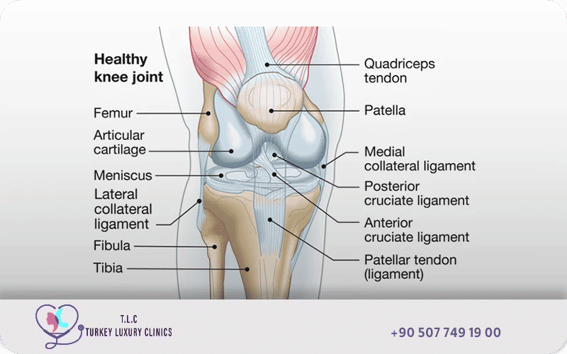
- Knee Joint Diagram
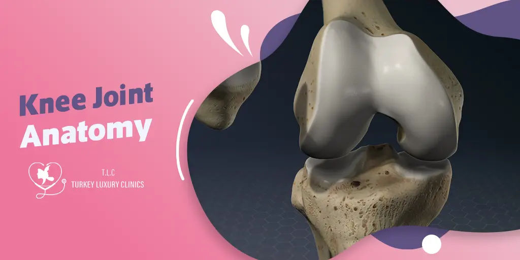
- Knee Joint Anatomy
- Knee Joint Articulations
- Bones of the Knee Joint
- Articular Cartilage
- Menisci
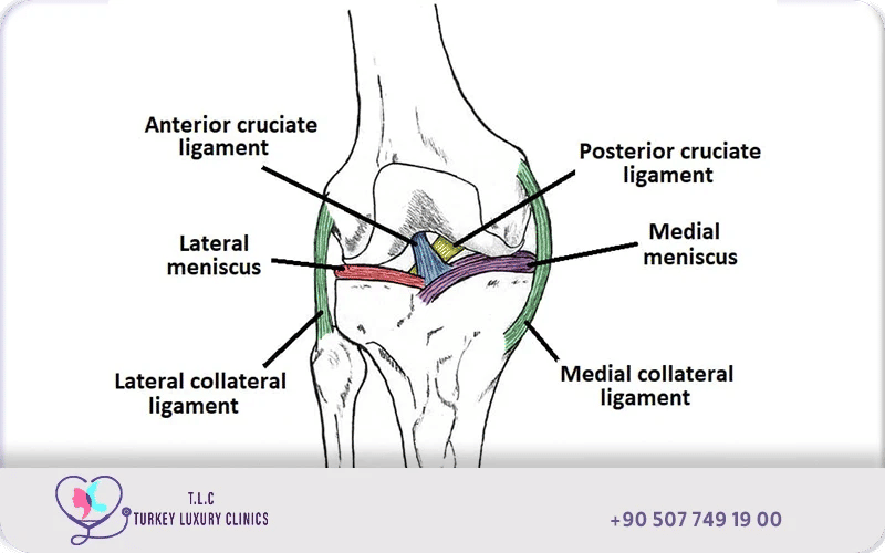
- Ligaments
- Muscles and Tendons
- Knee Joint Motions and Actions
- Knee Injuries and Diseases
- Knee Joint Imaging Techniques
- Knee Arthroscopy
- Knee Replacement Surgery
- New Innovations in Knee Replacement?
- Trusted Knee Replacement Surgery at Turkey Luxury Clinics
- FAQs About Knee Joint Anatomy
The knee is a modified hinge joint, classified as synovial, where the femur, tibia, and patella articulate to enable movement and support the body’s weight. Its primary movements are flexion (bending) and extension (straightening), ranging from 0° to approximately 150°, with limited rotational movement. These motions are essential for activities such as walking, running, and squatting, and are supported by ligaments including the ACL, PCL, MCL, and LCL.
The knee’s complex structure also allows slight medial and lateral rotation when bent, coordinated with patellar motion to distribute forces efficiently. Ligaments and surrounding muscles work together to limit excessive rotation and maintain stability, ensuring smooth, controlled motion while protecting the joint from stress and injury.
Knee disability can occur when any part of the knee joint anatomy is damaged, often due to knee arthritis or other degenerative conditions. In such cases, joint replacement procedures play a crucial role in relieving pain and restoring normal knee function.
Turkey Luxury Clinics guides you step by step—from understanding natural knee anatomy to exploring how a prosthetic knee can help you move smoothly, live pain-free, and regain a healthy, active life.
Knee Joint Diagram
The diagrams below illustrate the detailed anatomy of the knee joint, including bones, cartilage, ligaments, tendons, and muscles. These images help you visualize the relationships between each structure, understand joint movements, and see which areas may be affected by injury, arthritis, or other conditions. They are especially useful for explaining the surgical planning and placement of knee prosthetics.
Knee Joint Anatomy
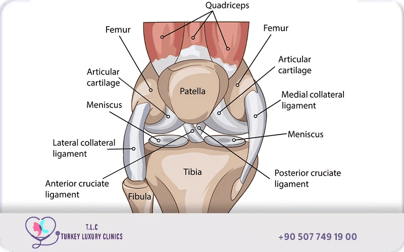
The knee is a complex synovial hinge joint connecting the femur, tibia, and patella (with the fibula nearby). It enables flexion/extension via the tibiofemoral and patellofemoral joints. Stability is provided by four major ligaments (ACL, PCL, MCL, LCL), with menisci acting as cushions.
The knee joint consists of two main articulations: the tibiofemoral joint, where the femur (thigh bone) meets the tibia (shin bone), and the patellofemoral joint, where the patella (kneecap) glides over the femur.
When looking at a knee joint picture anatomy, you will find a complex structure made up of bones, muscles, cartilage, ligaments, and tendons, all working together as an integrated system to ensure smooth movement, flexion, and stability.
Natural knee joint anatomy:
- Knee joint bones: Include the femur (thigh bone), tibia (shin bone), and patella (kneecap), which form the main articulating surfaces.
- knee joint anatomy muscles: Such as the quadriceps and hamstrings, which provide dynamic stability and assist in bending and extending the knee.
- Knee joint cartilage anatomy: The articular cartilage and menisci act as shock absorbers, reducing friction and protecting the bones from wear.
- Knee ligaments: The anterior cruciate ligament (ACL), posterior cruciate ligament (PCL), medial collateral ligament (MCL), and lateral collateral ligament (LCL) provide static stability and prevent abnormal movements.
- Knee tendons, Such as the quadriceps tendon and patellar tendon, connect muscles to bones and transmit the forces necessary for movement.
Knee Joint Articulations
The knee consists of two main articulations:
Tibiofemoral Joint
This is the primary articulation where the femoral condyles meet the tibial plateaus. It bears most of the body’s weight and allows flexion and extension.
Patellofemoral Joint
This articulation occurs between the patella and the femoral trochlea. The patella glides along this groove during knee movement, helping distribute forces across the joint.
Bones of the Knee Joint
Three main bones form the structural framework of the knee joint:
1. The femur, or thigh bone, forms the upper part of the joint and ends in rounded surfaces called the medial and lateral femoral condyles, which articulate with the tibia. These condyles allow the femur to roll and glide smoothly during knee movement.
2. The tibia, or shin bone, forms the lower weight-bearing surface of the joint. Its upper part contains two broad surfaces known as the tibial plateaus, which support the femoral condyles and transmit body weight from the thigh to the lower leg. Between these plateaus lies the intercondylar eminence, an elevated region that serves as an attachment point for the cruciate ligaments, which stabilize the joint.
3. The patella, or kneecap, is a triangular sesamoid bone embedded within the quadriceps tendon. Its main function is to improve the mechanical efficiency of knee extension by increasing the leverage of the quadriceps muscles. It also protects the front of the knee and helps distribute compressive forces during movement.
Articular Cartilage
The articular surfaces of the knee are covered by hyaline cartilage, a smooth and resilient tissue that allows the joint surfaces to move against each other with minimal friction. This cartilage acts as a protective layer that absorbs mechanical stress during weight-bearing activities such as walking or running.
Within this cartilage are specialized cells called chondrocytes, which maintain the extracellular matrix composed mainly of type II collagen and proteoglycans. These components give cartilage both strength and elasticity, allowing it to distribute loads evenly across the joint while maintaining flexibility.
Menisci
Between the femur and tibia lie two fibrocartilaginous structures known as the menisci. These structures, the medial and lateral meniscus, play a crucial role in maintaining knee stability and protecting the articular surfaces.
1. The medial meniscus has a semicircular shape and is firmly attached to the medial collateral ligament, which limits its mobility but increases joint stability.
2. In contrast, the lateral meniscus is more circular and slightly more mobile, allowing it to adapt more easily to the movement of the femoral condyles.
The menisci function primarily as shock absorbers, reducing the impact forces transmitted through the knee during weight-bearing activities. They also help distribute loads evenly across the joint, improve the congruence between the femur and tibia, and assist in the circulation of synovial fluid, which nourishes the cartilage.
Ligaments
Ligaments are strong connective tissues that link bones together and maintain knee joint stability by preventing excessive movement.
Collateral Ligaments, The MCL and LCL provide mediolateral stability, resisting valgus and varus stresses.
1. The medial collateral ligament (MCL) stabilizes the inner side of the knee and resists forces that push the joint inward, known as valgus stress.
2. On the outer side of the knee, the lateral collateral ligament (LCL) connects the lateral femur to the head of the fibula and resists various stresses, which pushes the knee outward.
Inside the joint are the cruciate ligaments, which cross each other and control the forward and backward movement of the tibia.
1. The anterior cruciate ligament (ACL) prevents the tibia from sliding forward relative to the femur and plays a major role in controlling rotational stability during dynamic activities.
2. The posterior cruciate ligament (PCL) prevents the tibia from moving backward relative to the femur and supports stability during deep knee flexion.
Both ligaments contain mechanoreceptors, specialized sensory structures that help the body detect joint position and movement, contributing to balance and coordination.
Muscles and Tendons
The muscles and tendons surrounding the knee generate the forces required for movement while also helping stabilize the joint.
1. The quadriceps muscles, located at the front of the thigh, are responsible for knee extension, allowing the leg to straighten during standing, walking, and climbing stairs.
2. Opposing them are the hamstring muscles, located at the back of the thigh, which flex the knee and allow the leg to bend during movement.
These muscles are connected to the bones through important tendons. The quadriceps tendon attaches the quadriceps muscles to the patella, transmitting muscular force during extension.
2. Below the patella, the patellar tendon connects the patella to the tibia and transfers the extension force to the lower leg, enabling powerful actions such as jumping and running.
Knee Joint Movement
Knee joint motions refer to the movements that occur at the knee, such as flexion, extension, and rotation, while knee actions describe the muscle groups responsible for producing these movements—for example, the quadriceps drive extension, and the hamstrings drive flexion.
The knee functions primarily as a synovial hinge joint, allowing flexion (bending) and extension (straightening) between the femur and tibia. Limited internal (medial) and external (lateral) rotation is possible, but only when the knee is flexed.
These movements are supported by gliding, rolling, and rotational mechanics, which are essential for smooth motion, joint stability, and efficient gait. The overall stability of the knee relies on the coordinated action of the joint capsule, menisci, ligaments, muscles, and the geometry of the femoral and tibial surfaces. Together, these structures enable controlled, stable movement while protecting the joint from excessive forces during weight-bearing activities.
Knee Joint Movement Types
Action | Primary Movement | Primary Muscles |
Flexion | Bending the knee (heel to glute) | Hamstrings |
Extension | Straightening the knee | Quadriceps |
Internal Rotation | Turning the tibia inward (knee flexed) | Popliteus, Semitendinosus |
External Rotation | Turning the tibia outward (knee flexed) | Biceps Femoris |
Primary Knee Movements
The primary movements of the knee are flexion, extension, and rotation, which represent the three main types of knee movement. The knee joint primarily functions as a hinge joint, allowing flexion and extension with limited rotation when flexed.
Flexion occurs when the hamstrings pull the lower leg backward toward the thigh, allowing approximately 140°–160° of motion, while extension, driven by the quadriceps, straightens the leg. A "screw-home" mechanism occurs at full extension for stability, involving subtle rotation.
Complex Knee Movements
Complex knee movements involve coordinated mechanical actions between the femur, tibia, patella, ligaments, and surrounding muscles to maintain joint stability and smooth motion.
Complex Movement | Mechanical Description | Functional Purpose |
Screw-Home | Slight external rotation of the tibia at full extension | Locks the knee in a stable position for standing |
Unlocking (Popliteus) | Internal rotation of the tibia to start flexion | Releases the knee from the locked position for bending |
Roll & Glide | Simultaneous rolling and sliding of the femur on the tibia | Prevents the femur from sliding off during deep flexion |
Patellar Tracking | Patella moves up and down in its groove | Increases quadriceps leverage and distributes forces across the joint |
Knee Joint Biomechanics
Knee joint biomechanics involves the complex interaction of two main joints, the tibiofemoral and patellofemoral joints, to facilitate movement while maintaining stability under heavy loads.
Flexion is the bending of the leg, bringing the calf toward the back of the thigh. This movement is mainly performed by the hamstrings (biceps femoris, semitendinosus, semimembranosus) with assistance from the popliteus, gracilis, and sartorius, allowing about 140°–160° of motion.
Extension straightens the leg from a bent position and is primarily controlled by the quadriceps femoris, reaching 0° neutral up to 5–10° hyperextension.
When the knee is flexed, it allows limited rotation. Medial (internal) rotation turns the leg inward, powered by the popliteus, semimembranosus, and semitendinosus, while lateral (external) rotation turns the leg outward, mainly driven by the biceps femoris. Typical rotation ranges are ~10° internally and 30°–40° externally.
A key biomechanical feature is the screw-home mechanism, where at full extension, the tibia rotates slightly externally (or the femur medially), locking the knee into a stable, weight-bearing position. The popliteus muscle then unlocks the joint to initiate flexion.
Additionally, the knee exhibits roll-and-glide motion as the femur moves over the tibia, a dynamic rotation axis that shifts with bending, and patellar motion that improves quadriceps leverage and distributes load across the joint. Proper coordination of these structures ensures smooth, stable knee mechanics and efficient gait.
Typical Knee Joint Movement Ranges
Movement | Typical Range |
Flexion | 120°–150° |
Extension | 0° to 5–10° hyperextension |
Internal Rotation | ~10° (when flexed) |
External Rotation | ~30°–40° (when flexed) |
Knee Joint Motions and Actions
Main knee actions include stabilizing the body during walking, running, and jumping, with the quadriceps extending and hamstrings flexing the leg. Knee joint motions involve coordinated action of muscles, ligaments, and articular surfaces.
For example:
- Quadriceps muscles perform knee extension
- Hamstring muscles perform knee flexion
- Popliteus muscle assists in unlocking the knee
Load and Gait Mechanics of Knee
The knee joint is subjected to significant mechanical forces during daily activities. The mechanical axis of the lower limb runs from the femoral head to the ankle and normally passes through the center of the knee. The anatomical axis between the femur and tibia typically forms a valgus angle of about 172°–177°. Deviations from this alignment can lead to varus or valgus knee deformities, increasing stress on joint surfaces and accelerating the development of osteoarthritis. During walking, the knee may experience forces ranging from three to seven times body weight.
Knee Injuries and Diseases
Several common knee disorders can lead to severe damage of the knee joint anatomy, often making knee replacement the most effective treatment option. The most frequent causes include:
- Osteoarthritis: Age-related cartilage wear leading to pain, stiffness, and bone-on-bone friction.
- Rheumatoid arthritis: Chronic inflammation that progressively destroys cartilage and soft tissues.
- Post-traumatic arthritis: Damage caused by previous fractures, ligament tears, or meniscus injuries.
- Advanced meniscus or cartilage loss: Severe deterioration that can no longer be repaired through arthroscopy or conservative treatments.
Anatomy of the knee joint diagrams are essential for understanding which part of the knee is damaged and for planning the right treatment approach, such as knee replacement surgery.
Knee joint replacement involves removing the damaged cartilage and bone surfaces of the femur (thigh bone), tibia (shin bone), and sometimes the patella (kneecap), and replacing them with artificial components made of metal and medical-grade plastic.
Knee Joint Imaging Techniques
Getting the anatomy of knee joint images is essential for diagnosing knee joint conditions and planning replacement surgeries. The main imaging techniques include:
- knee joint anatomy X-ray
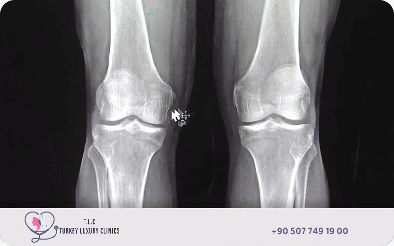
A knee X-ray is a diagnostic imaging test that produces black-and-white images of the knee joint, mainly highlighting the bones — such as the femur, tibia, and patella. It helps detect fractures, joint space narrowing, and alignment issues. Although X-rays provide limited details on soft tissues like ligaments or cartilage, they are often the first diagnostic step for knee pain or injury.
Orthopedic surgeons usually request a knee X-ray for diagnosis of the primary cause, before knee replacement surgery and after the surgery for follow-ups.
- Knee joint MRI anatomy:
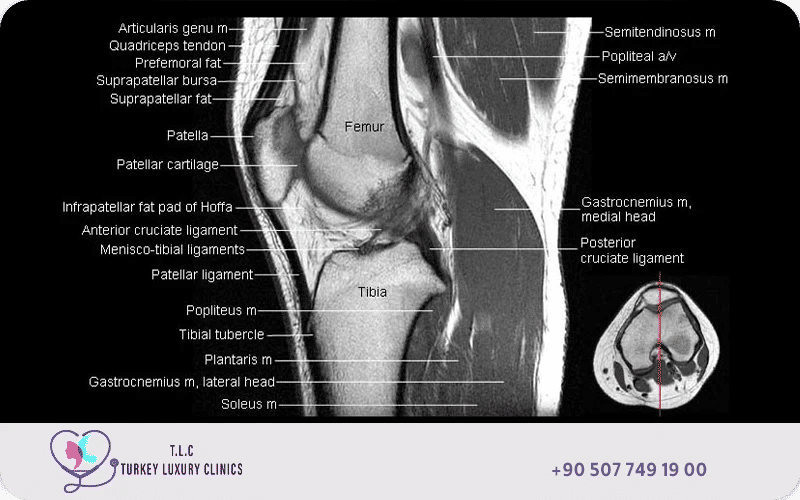
Knee joint MRI anatomy refers to the detailed visualization of the structures within the knee using magnetic resonance imaging, to create cross-sectional images of the bones, cartilage, ligaments, tendons, muscles, and nerves in and around the knee joint.
It is commonly used before surgery for accurate diagnosis and surgical planning, especially in complex cases where soft tissue damage must be evaluated.
- Knee joint anatomy ultrasound :
Knee joint ultrasound is a non-invasive imaging technique that uses sound waves to visualize the superficial soft tissues of the knee, including tendons, ligaments, muscles, and bursae. It is excellent for evaluating extra-articular structures and some periarticular pathology like fluid collections and synovitis
Ultrasound is useful for real-time assessment of tendons, ligaments, and fluid accumulation around the joint. It is often used during diagnosis or post-surgery follow-up to detect fluid buildup or inflammation.
- Knee joint anatomy 3D image:
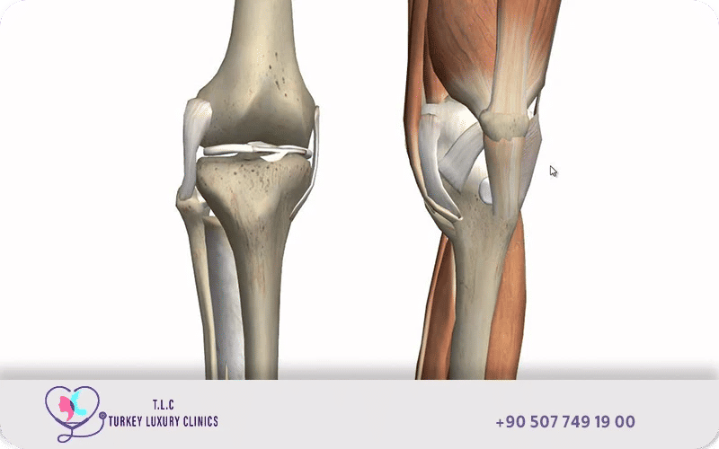
3D knee imaging, primarily utilizing 3D MRI and sometimes combined with CT, offers enhanced visualization and analysis of the knee joint compared to conventional 2D imaging
Knee joint 3D image is a crucial component of personalized knee replacement surgery, enabling the creation of custom-fit implants and surgical guides for improved accuracy, better patient outcomes, and potentially faster recovery.
Knee Arthroscopy
Knee arthroscopy in Turkey is a minimally invasive surgical procedure used to diagnose and treat various knee problems, including issues related to ligaments and tendons.
As an advanced technique, it involves inserting an arthroscope—a small camera—through tiny incisions, usually 1 to 2 cm, to visualize the inside of the knee joint allowing surgeons to accurately repair or remove damaged tissues with minimal trauma and faster recovery.
Knee Replacement Surgery
Knee joint replacement, also known as knee arthroplasty, is a surgical procedure in which the damaged or worn-out parts of the knee joint are replaced with an artificial prosthetic implant
This operation is primarily performed to relieve chronic pain, restore joint function, and improve mobility in cases of severe osteoarthritis or other degenerative conditions where non-surgical treatments have failed.
Knee replacement offers several benefits, including up to 90–95% pain relief, improved joint function, restored mobility, and better quality of life with enhanced knee flexion and stability.
The procedure boasts a success rate of 90–95%, with most patients experiencing substantial pain relief and improved mobility within a recovery period of 6 to 12 months.
In Turkey, Knee replacement surgery is a popular and cost-effective option for patients seeking treatment for severe knee problems.
Read more about: Top indications for knee replacement, When is surgery necessary, and who is a candidate for knee replacement
New Innovations in Knee Replacement?
Turkey is at the forefront of advanced medical technologies, offering cutting-edge innovations that improve the accuracy, safety, and long-term outcomes of knee replacement surgeries. One of the most notable advancements is the robotic-assisted knee replacement.
Robotic-assisted knee replacement is a modern surgical technique in which a robotic arm assists the surgeon during the procedure.
The surgeon remains in full control, but the robotic system enhances precision by guiding accurate bone cuts, optimizing implant positioning, and ensuring better alignment.
This advanced approach often leads to a joint that feels more natural and can result in faster recovery, less postoperative pain, and improved long-term function.
Trusted Knee Replacement Surgery at Turkey Luxury Clinics
At Turkey Luxury Clinics, we provide expert knee replacement surgeries tailored to each patient’s needs. With advanced surgical techniques, experienced orthopedic surgeons, and world-class facilities, we ensure a smooth treatment journey and long-lasting results. Contact us today to book your consultation and take the first step toward a pain-free life.